

Research
New Center Offers a ‘Look’ inside the Brain
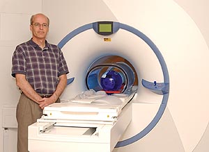 |
| MAGNETIC QUALITY: Richard Aslin, professor of brain and cognitive sciences and director of the Center for Brain Imaging, leads an interdisciplinary team using the new fMRI system to explore how the human brain functions. The center will be formally dedicated this fall. |
Rochester scientists have a powerful new tool for understanding how the human brain manages such complicated tasks as processing language and speech, learning new skills, and how it handles other neurological and sensory processes that have largely remained mysterious.
Formally dedicated this fall, the Rochester Center for Brain Imaging features a state-of-the-art magnetic resonance imaging system that can pinpoint which parts of the brain are active while a variety of tasks are being performed.
Based on technology that has been used in hospitals as a noninvasive diagnostic tool for the past decade or so, the new machine has twice the magnetic field strength of standard clinical MRIs. It’s designed to indicate not just anatomical details but which parts of the brain are active while the brain is functioning, a system known as functional magnetic resonance imaging, or fMRI. Only about 30 such fMRIs exist in the nation.
“The beauty of this is that it will allow us to ‘see’ inside the brains of people in a way that’s noninvasive and in a way that we know is very, very safe,” says Richard Aslin, the University’s William R. Kenan Jr. Professor and director of the center.
Established through grants from the National Science Foundation, the David and Lucille Packard Foundation, and the Schmitt Program on Integrative Brain Research, the center is housed in renovated space on the River Campus just across Elmwood Avenue from the Medical Center.
Researchers representing fields that include brain and cognitive sciences, biomedical engineering, electrical and computer engineering, neurology, pediatrics, ophthalmology, radiology, cardiology, and clinical and social sciences in psychology have expressed interest in using the new center.
The center also will train undergraduates, graduate students, and postdoctoral fellows as they learn how to use MRI in their work.
“This center is a wonderful addition to the research tools for a widespread and important group of scholars at the University,” says Provost Charles E. Phelps. “It significantly augments our research ability in both the College and the Medical Center.”
Finding a way to watch the human brain in action has long been a goal of cognitive and medical scientists. In the past, researchers have gathered information about the mind by observing how people respond to external stimuli.
The fMRI system, on the other hand, offers a glimpse of which parts of the brain are active during tasks by measuring changes in a magnetic field created by the system’s powerful magnet. Those changes occur as blood flows to active areas of the brain. By analyzing the changes, researchers can put together pictures of how the brain works.
“Functional MRI is an area of research that is growing exponentially,” Aslin says.
In the center, test subjects will lie on their backs with their heads resting in a 40-centimeter “sweet spot” while the 3.0 Tesla magnet pulsates around them. Participants will be able to see a screen projected above their heads, or listen to sounds over headphones, and be asked to manipulate a small set of handheld controls in response to questions about what they see or hear.
Projects slated for the new center include one led by Daphne Bavelier, associate professor of brain and cognitive sciences, which investigates how the brain is reorganized when an entire sense—such as hearing in deaf individuals—is absent.
Aslin and Elissa Newport, professor and chair of brain and cognitive sciences and the George Eastman Professor of Brain and Cognitive Sciences, will search for areas of the brain that are activated as adults and children rapidly learn new skills, including novel miniature languages.
A team led by Bavelier, Newport, and Ted Supalla, associate professor of brain and cognitive sciences, will investigate how language is represented in the brain by studying individuals who use sign language rather than spoken language.
Jianhui Zhong, professor in the Departments of Radiology, Biomedical Engineering,
and Physics, and Kevin Parker, dean of the School of Engineering and Applied
Sciences, plan to develop new techniques for gathering images from the magnet
and visualizing their three-dimensional properties.
Pilot studies with the new system got under way this summer.
