

A MAG-nificent Specimen
Curators from the Memorial Art Gallery and scientists from the Medical Center team up to study a 2,000-year-old mummy. By Scott Hauser
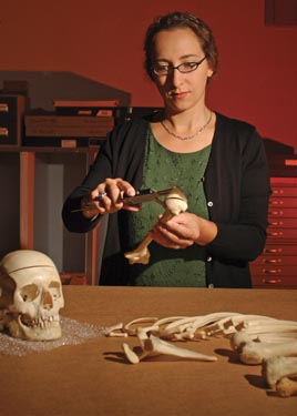 |
| ANTHROPOLOGIST: Jennifer Prutsman-Pfeiffer, a forensic anthropologist and pathologists’ assistant at the Medical Center, found no evidence that the man whose remains were mummified sometime between 30 C.E. and 330 C.E. had died of anything but natural causes. |
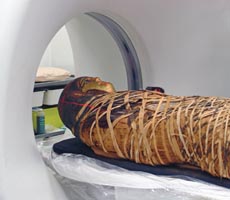
Like all good forensic mystery stories, this one starts in the predawn hours on a fall day suspiciously close to Halloween. At about 4 o’clock on a late September morning, a mummy was carried out of the Memorial Art Gallery and slid gently into the back of a funeral home’s van.
As the modern-day hearse headed toward Strong Memorial Hospital, those who saw the body—its face covered by a golden mask—speculated with nervous excitement about what the approaching investigation might reveal.
Who’s behind the gilded faceplate? How did he live? How did he die?
And, most important, how could the body best be explained to the public?
Once at Strong, the story moves quickly through centuries of archaeological science and anthropological research.
The early morning appointment was part of an unusual cross-University collaboration involving curators from the gallery and scientists from the Medical Center. Using a state-of-the-art computed tomography (CT) imaging system, as well as the latest in forensic science and forensic art, the team hoped to uncover clues to some of the secrets of life and death in ancient Egypt.
The mummy meets the 21st century, so to speak.
“It’s fascinating to me that I can take everything I know forensically and apply it to someone who is 2,000 years old,” says Jennifer Prutsman-Pfeiffer, a forensic anthropologist at the Medical Center, who was part of the project.
Prutsman-Pfeiffer, who serves as a consultant for medical examiners in 10 upstate and western New York counties, is a pathologists’ assistant in the Department of Pathology and Laboratory Medicine at Strong. As a trained forensic anthropologist, she’s often asked to help piece together the puzzles of mysterious deaths.
And the case of the gallery’s mummy certainly qualified, she says.
“I had to stop a couple times and think, ‘Wow, this guy is 2,000 years old.’”
The “guy”—and one of the project’s findings confirmed that the body is that of a male—now is part of the gallery’s exhibition Protected for Eternity: The Coffins of Pa-debehu-Aset, which opened in October 2003 and is expected to stay on display through 2007. The mummy, on loan from the Peabody Essex Museum in Salem, Massachusetts, is helping round out a picture of the religious, cultural, and social practices of people who lived near the Nile more than two millennia ago.
Centered on two ornately decorated coffins of a middle-class Egyptian named Pa-debehu-Aset, who lived 2,300 years ago, the exhibition guides visitors through the process of mummification and explains its role in the ancient Egyptian belief system. The exhibit features canopic jars—used to store the internal organs of the dead—amulets, and other funerary objects. There also are interactive stations that explain mummification and explain the work of conservators to preserve ancient artifacts.
Considered one of the most significant acquisitions in the museum’s history when they were purchased in 2000, the coffins had long been separated from the body of their owner.
Nancy Norwood, the curator for the 750-square-foot exhibit, says including a mummy, even one from a different period of history from the coffins, adds an important educational aspect to the exhibition.
“We wanted to emphasize that there’s a strong spiritual reason behind mummification,” Norwood says. “[A mummy] really does emphasize the humanity of the process.”
Thanks to the new Rochester work, Norwood and other researchers also have a little better picture of the humanity of the person whose remains were mummified sometime during the Roman era (30 C.E. to 330 C.E.) in ancient Egypt.
 |
 |
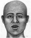 |
| ‘PATIENT’: Using data collected by Strong’s CAT imaging system, forensic artist Kristin Davies created a portrait of what the Egyptian man may have looked like 2,000 years ago. | ||
The analysis shows that he stood about 5 feet, 6 inches tall and was between the ages of 20 and 30 years old when he died. His teeth were in excellent condition, indicating that he probably enjoyed a somewhat comfortable upper middle-class lifestyle. And the condition of his bones gives no indication that he died a violent death.
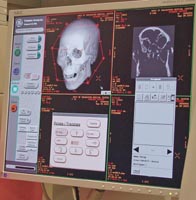
That information adds to earlier studies of the mummy, but the new scans—showing anatomical details at the level of millimeters—have allowed researchers to put together a highly detailed digital image of the body and, in particular, the skull. From those images, an FBI-trained forensic artist has drawn a portrait of what the man may have looked like.
“The potential for gaining new scientific knowledge about the mummy was very exciting, considering the collaborative effort that could take place between the gallery and Strong,” Norwood says.
Trying to understand the process of mummification—along with its religious and spiritual underpinnings—has fascinated observers since at least 450 B.C.E., when the Greek historian Herodotus recorded what’s considered the earliest existing account of mummification in Egypt. But it wasn’t until the late 1800s and early 1900s that interest in ancient Egypt and its antiquities really took off in the public imagination as expeditions began to discover the tombs of pharaohs, including the famous 1923 excavation of the tomb of Tutankhamen.
But those late 19th-century efforts often faced a dilemma: To find out who was wrapped in the layers of linen, researchers (and sometimes less-than-scrupulous collectors) had to unwrap the bodies, a process that often destroyed the materials under study.
John Taylor, a curator for a British Museum exhibition that’s based on the most thorough CT scanning effort of a mummy yet undertaken, says Egyptologists were quick to realize that imaging technology could provide a way to “see” inside mummies.
Archaeologists at the turn of the 20th century were among the first to use X-rays, Taylor says, and X-rays remained the tool of choice until the 1980s, when CT scanning became widely available.
“For the past 50 years, [unwrapping] has hardly ever been done,” Taylor says. “What’s nice about CT scanning is that it preserves everything in its original context.”
That context was exactly what Norwood was looking for as she organized the exhibition in the gallery’s Gill Discovery Center.
“The potential for gaining new scientific knowledge about the mummy was very exciting, considering the collaborative effort that could take place between the gallery and Strong.” —Nancy Norwood, curator at the Memorial Art Gallery
Norwood turned to Mimi Leveque, a freelance conservator who had helped restore the Pa-debehu-Aset coffins. Leveque recently had conserved the Peabody Essex mummy, including restoring the luster to the golden mask that represents his face, and she helped arrange for the mummy to be on loan to the gallery.
Leveque, who has collaborated in several conservations of mummies around the country, says using CT scanning to study mummies is not unusual, but it’s not something that many museums can carry out.
She says the technology can be helpful in understanding not just the person who was mummified, but also the era in which the mummification took place. During the process, priests often placed amulets and other artifacts in the wrappings to help protect the person on the journey to what Egyptians believed was the afterlife.
Those kinds of details about funerary practices can help pinpoint time periods in the history of mummification.
“It’s possible to get a fairly clear idea about the person who is inside,” Leveque says. “But it’s also luck of the draw when you do a scan as to what you’re going to see.”
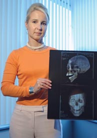 |
| ARTIST: Kristin Davies, an FBI-trained forensic artist
for the Rochester Police Department, helped make sure the new scans included information she would need to create a likeness of the mumified man. |
Rochester researchers had some idea of what they would see in the Peabody Essex mummy because it had been CT’d earlier in Massachusetts, and it arrived in Rochester with a copy of the scans. But the images had never been analyzed.
As Norwood turned to medical experts in Rochester and at the University, she discovered that, even though the images were less than five years old, they were, by the standards of technology, out-of-date.
“The real benefit to scanning the mummy was that the technology has improved so much in just a few years,” Norwood says.
Another advantage was that new images would allow researchers to orient the mummy’s face digitally to provide a head-on view of the skull. Earlier scans were unable to capture that perspective because the mummy’s head is out of alignment with his body. And moving the head to align with the scanner would damage the mummy.
That head-on view—known scientifically as the “Frankfurt horizontal”—is important because the perspective is used by forensic artists to re-create facial characteristics when the only information available about an unknown person is a skull. Prutsman-Pfeiffer contacted Kristin Davies, a forensic artist for the Rochester Police Department with whom she often collaborates on missing person cases, to see if Davies thought she could create a likeness of a 2,000-year-old missing person whose skull would be visible only on a computer screen.
“I thought it was fascinating,” Davies says. “It was a once-in-a-lifetime opportunity.”
And so an appointment was made in September 2003 at the Department of Radiology at the Medical Center, where Norwood, Prutsman-Pfeiffer, Davies, and Leveque joined Patrick Fultz, associate professor of radiology and the director of body imaging, along with several other technicians and researchers, to scan the mummy.
Scheduled for 5 a.m. to avoid interfering with other appointments, the mummy was treated as if it were a living patient. And given his condition he was just that—patient. While a live human being can rarely stay still for an extended scan, the mummy was able to be analyzed in a 90-minute session.
As the body lay on the table of the CT scanner, the machine’s powerful system of X-ray beams captured a series of images of the body at intervals of just a few millimeters. That information was fed into a computer to create a three-dimensional, computerized image of the skull.
Davies, who helped direct the effort to make sure the images included the critical Frankfurt horizontal perspective of the skull, then used a complex method of correlating measurements of bone structures with facial tissues and characteristics to create a drawing of what the Egyptian man may have looked like.
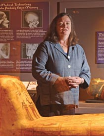 |
| CURATOR: Gallery curator Nancy Norwood, says the mummy helps emphasize the spiritual basis of ancient Egyptian beliefs about the afterlife. |
The drawing shows the face of a man who could have a living counterpart in the Egypt of 2004. That’s not unexpected, given that 2,000 years is a relative blink of the eye compared to the history of human evolution, Davies says.
“I was drawing somebody who could be alive today,” Davies says.
Prutsman-Pfeiffer, who helped analyze the scans, was able to provide more context about the body, and was able to confirm details about his age and height. The body also has several broken bones, but the fractures appear to have occurred after he died, either during the embalming process or while he was first shipped to the United States sometime in the late 1800s or early 1900s.
The new scans provide a source for further research in the use of modern medical technology to study ancient peoples, says Prutsman-Pfeiffer.
“It would have been better if we had more of a biological profile to work with,” she says. “But like anything academic, this has great value to the community.”
Norwood, who incorporates some of the older Peabody Essex images in the exhibition as a way to show visitors how technology augments the study of ancient societies, hopes to continue to explore ways to use the new set of digital images taken at Rochester for other projects.
She would like to create a “virtual mummy” that visitors could manipulate on a computer, allowing people to see the different layers—from the outer wrappings to the bones—that are hidden beneath the mummy’s tightly wound resin-coated linen strips.
“This is not a static exhibition,” Norwood says. “This project will continue to evolve.”
Scott Hauser is editor of Rochester Review.
