In Review
 BRAIN BIOMARKER: A new test approved by the FDA and based on Rochester research can diagnose brain injuries from a blood sample. (Photo: Adobe Stock)
For the first time in the US, a blood test will be available to help doctors determine if people who’ve experienced a blow to the head could have a traumatic brain injury such as brain bleeding or bruising. Until now, physicians have relied on subjective markers—mainly patient-reported symptoms such as headaches, nausea, or light sensitivity—to make an educated guess as to which individuals have brain trauma and require a head CT scan.
The new test, called the Banyan Brain Trauma Indicator, provides an objective measure of injury that can be obtained quickly and easily in busy emergency departments. The US Food and Drug Administration approved the test last February as part of a fast-track program to get breakthrough technologies to patients more quickly. The major study that led to approval of the test was published in the Lancet Neurology.
“Many concussion patients don’t seek medical care for their injury, a decision due in part to the perception that emergency departments have nothing to offer in terms of diagnosis,” says lead study author Jeffrey Bazarian, a professor of emergency medicine at the Medical Center. “The results of this study show that we now have something to offer—a brain biomarker blood test. The ability of this test to predict traumatic injuries on head CT scans will soon allow emergency physicians to provide patients with an unbiased report on the status of their brain.”
The test detects two brain proteins that are present in the blood soon after a hit to the head. The study shows that if the test is negative—meaning that the brain proteins aren’t present—it is highly unlikely that a traumatic intracranial injury exists, and a head CT scan can be safely avoided. If the test is positive, a brain injury may be present and the patient should receive a head CT scan to further assess the damage and guide treatment.
—Emily Boynton
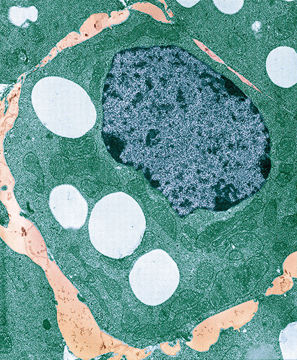 THE LURE OF LIPIDS: Lipid droplets—microscopic pockets of lipids inside cells—are a “hot area of research.” (Photo: University of Edinburgh/Wellcome Collection)
Lipid Droplets Play Role in Gene Expression
Lipid droplets—microscopic pockets of lipids located inside cells—are “a really hot area of research,” says Michael Welte, professor and chair of biology at Rochester. That’s because they have been found to play a role in gene expression. A study led by Welte and published in the journal eLife describes how lipid droplets fulfill that role.
Welte used fruit fly embryos to study how lipid droplets influence a set of proteins called histones. If there are too few histones, genes might be expressed that shouldn’t be. Too many histones can cause cells to have trouble dividing their chromosomes.
Welte discovered that lipid droplets play an important role in regulating a particular histone called H2Av. Acting like pacemakers, the lipid droplets regulate how fast H2Av enters a cell’s nucleus by storing the histone until the nucleus needs it.
Identifying the functions of lipid droplets gives researchers insight into how embryos develop and survive: without lipid droplets regulating H2Av, embryos can become compromised.
The findings could cause researchers to reconsider how they look at lipid-related diseases. Lipid droplets are dysfunctional in disease states like obesity (too many lipid droplets) or lipodystrophies (too few). “The cause of these diseases—too much or too little fat—has to do with how much lipid you have,” Welte says. “Our work suggests that when looking at these disease states, people also need to look at what happens to the proteins, because these lipid droplets have this second function beyond handling fat.”
—Lindsey Valich
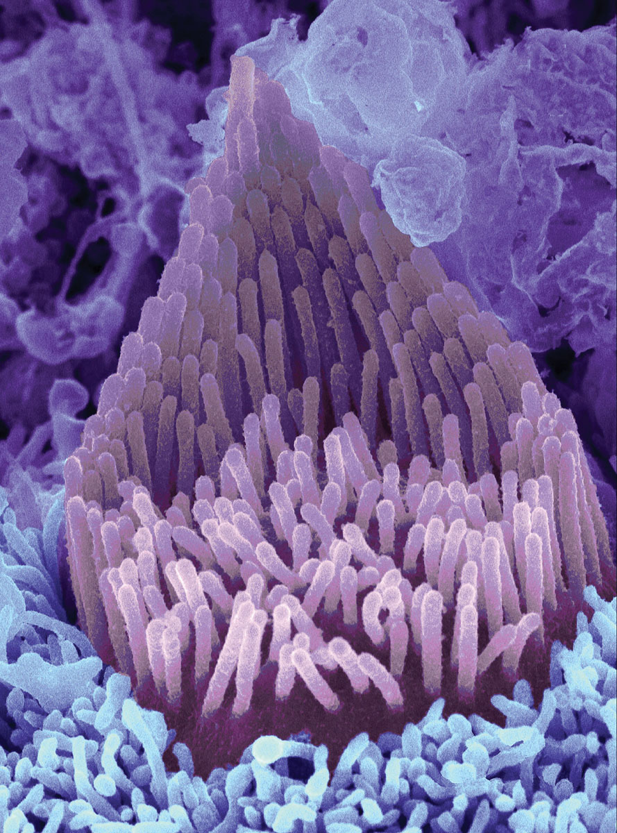 HAIRS THAT ‘HEAR’: Researchers have regrown hair cells in the cochlea, where sound vibrations are converted into electrical signals, allowing people to hear. (Photo: David Furness/Wellcome Collection)
New Therapy for Hearing Loss?
Researchers have taken an important step toward what may become a new approach to restore hearing loss. In a study published in the European Journal of Neuroscience, scientists have been able to regrow sensory hair cells found in the cochlea, a part of the inner ear that converts sound vibrations into electrical signals and can be permanently damaged with age or from noise.
“It’s funny, but mammals are the oddballs in the animal kingdom when it comes to cochlear regeneration,” says Jingyuan Zhang, a postdoctoral associate in neuroscience and the first author of the study. “We’re the only vertebrates that can’t do it.”
In a previous study, Patricia White, a research associate professor at the Del Monte Institute for Neuroscience, identified a family of receptors—called epidermal growth factor (EGF)—responsible for activating support cells in the auditory organs of birds. When triggered, the cells proliferate and foster the generation of new sensory hair cells. She speculated that the signaling pathway could be manipulated to produce a similar result in mammals.
In the new study, she tested that theory, focusing on a specific receptor called ERBB2, which is found in cochlear support cells. White and her team found that activating the ERBB2 pathway led to a proliferation of cochlear support cells, which in turn activated neighboring stem cells to become new sensory hair cells. The process not only could lead to the regeneration of sensory hair cells, but also could support their integration with nerve cells.
The process of repairing hearing is complex, says White. “You have to regenerate sensory hair cells and these cells have to function properly and connect with the necessary network of neurons.” But her research “could represent a new approach to cochlear regeneration and, ultimately, restoration of hearing.”
—Samantha Jean
he Rising Rates of End-of-Life Rehab
A new study indicates a growing trend of potentially unnecessary—and harmful—high-intensity rehabilitation services for residents of nursing homes. The study finds that the trend is on the rise for patients in the last 30 days of life, and is especially concentrated in the last seven days.
“This study raises several concerns and questions regarding the scope and intensity of therapy provided to nursing home residents prior to death,” says Helena Temkin-Greener, a professor in the Medical Center’s public health sciences department and lead author of the study.
The researchers note that financial considerations could play a role in the trend. Nursing home Medicare reimbursement rates are based on the complexity, intensity, and amount of staff time dedicated to care. Patients who receive high levels of rehabilitation services make such facilities eligible for the highest level of reimbursement.
Temkin-Greener and her colleagues analyzed data from 647 nursing homes in New York state. Specifically, they focused on residents who had received very high to ultrahigh rehabilitation services during the last 30 days of life. They found that the number of residents receiving ultrahigh rehabilitation increased by 65 percent between 2012 and 2015 and that most of the rehabilitation therapy residents received was concentrated in the last seven days of life. They also found a significantly higher use of these services in for-profit nursing homes than in nonprofit homes.
“These are often sick and frail patients in whom the risks of intensive levels of rehabilitation actually outweigh the benefits,” says Thomas Caprio, a geriatrician at the Medical Center and coauthor of the study.
Temkin-Greener says that if the expansion of the services is “being driven by a failure to recognize that a resident is approaching the end of life, then it calls for improving the skills of nursing home teams. If it is being driven by financial considerations, then regulatory and policy interventions may be necessary.”
The research appears in the Journal of the American Medical Directors Association.
—Mark Michaud
Rochester Review
Fall 2018
Vol. 81, No. 1

Section as a PDF