Society & Culture
The cookbook that taught generations of Jews how to become American
A new book reveals how The Settlement Cook Book became an unlikely guide to immigration, assimilation, memory, and belonging.

URochester students gain hands-on experience designing, building, and racing off-road vehicles together.


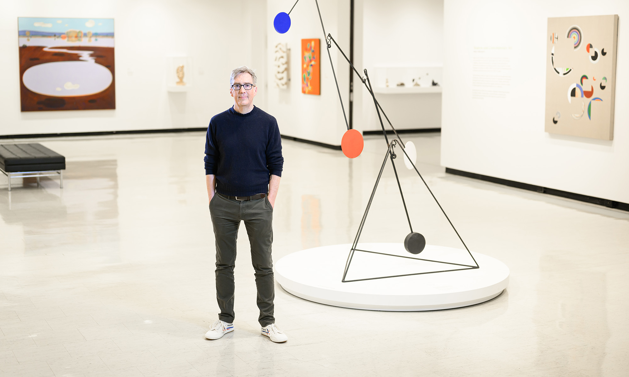

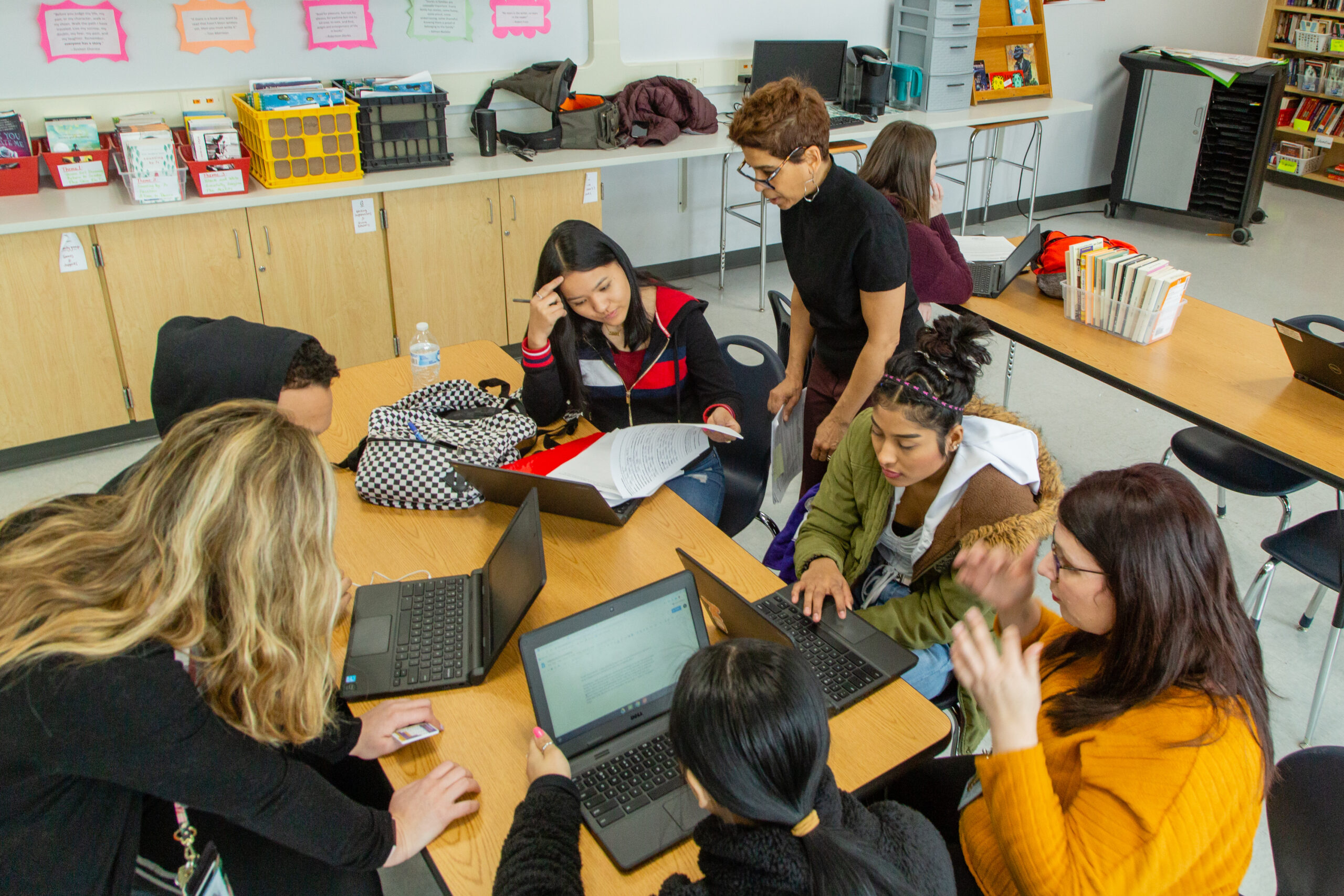


At the University of Rochester, there’s no need to “pick a lane.” Here, art meets analytics, science meets soul, and curiosity leads to unexpected outcomes. It’s a home where thinkers and doers, scholars and starters, healers and leaders come together to create a world that’s ever better.