|
|
|
|
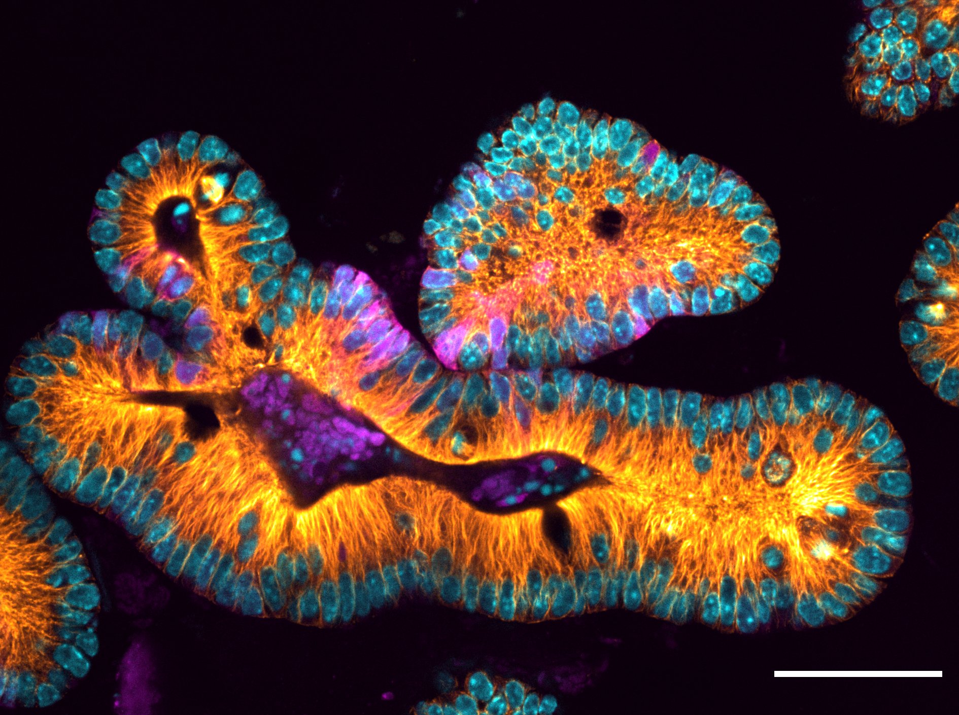 University of Rochester biologist Dan Bergstralh studies organoids—tissue cultures that replicate the complexity of an organ (such as the one pictured)—to determine how organs form and develop. A new light sheet microscopy resource to be constructed on campus will allow researchers like Bergstralh to study biological processes in groundbreaking ways by collecting and analyzing 3-D images of complex cellular structures. (Photo by Nicole Dawney)
Grant funds advance in biological imagingA new multidisciplinary collaboration between the departments of biology and biomedical engineering, the Institute of Optics, and the Goergen Institute for Data Science will develop and build a novel light-sheet microscope that employs freeform optical designs devised at Rochester, allowing for cutting-edge scientific research in biological imaging.
Michael Welte, professor and chair of the Department of Biology, is the lead principal investigator of the project, which was awarded a $1.2 million grant from the Arnold and Mabel Beckman Foundation.
The microscope, which will be housed in a shared imaging facility in Goergen Hall and is expected to be online in 2022, enables three-dimensional imaging of complex cellular structures in living samples. Researchers and engineers will continually improve the microscope, and it will eventually become a resource for the entire campus research community.
While many other research microscopes illuminate objects pixel by pixel, light-sheet technology illuminates an entire plane at once. The result is faster imaging with less damage to samples, enabling researchers to study biological processes in ways previously out of reach. Read more here.
Research sheds light on vision loss in Batten DiseaseProgressive vision loss, and eventually blindness, are the hallmarks of juvenile neuronal ceroid lipofuscinosis (JNCL) or CLN3-Batten disease. New research led by Ruchira Singh, an associate professor in the Department of Ophthalmology and Center for Visual Science, shows how a mutation associated with the disease potentially leads to degeneration of light sensing photoreceptor cells in the retina, and subsequent vision loss. The study appears in the journal Communications Biology.
Batten disease is caused by a mutation in the CLN3 gene, which is found on chromosome 16. Most children suffering from JNCL have a missing part in the gene which inhibits the production of certain proteins. Rapidly progressive vision loss can start in children as young as 4, who eventually go on to develop learning and behavior problems, slow cognitive decline, seizures, and loss of motor control. Most patients with the disease die between the ages of 15 and 30.
To study Batten disease in patient’s own cells, the research team reengineered skin cells from patients and unaffected family members to create human-induced pluripotent stem cells. These cells, in turn, were used to create retinal cells which possessed the CLN3 mutation. Using this new human cell model of the disease, the new study shows for the first time that proper function of CLN3 is necessary for retinal pigment epithelium (RPE) cell structure. RPE is the cell layer in the retina that nourishes light sensing photoreceptor cells in the retina and is critical for their survival and function and thereby vision.
Understanding how RPE cell dysfunction contributes to photoreceptor cell loss in Batten disease will enable researchers to target specific cell types in the eye using potential future gene therapies, cell transplantation, and drug-based interventions.
Read more here.
NIH grant writing webinar for junior investigators
| A webinar hosted by the National Research Mentoring Network (NRMN) from noon to 2 p.m. Tuesday, March 16 will address best practices for effective grantsmanship, approaches for competitive applications, common mistakes made by junior investigators and tips to increase chances for success. |
The focus will be on NIH’s research grants, especially the common R01 funding mechanism.
Topics will include:
- key required application components
- specific content that should be included when completing the individual sections
- NIH’s research grant review process, scoring system, and review criteria
Keeping abreast of the University's response to COVID-19
Here are important links for researchers:
PLEASE NOTE that the University’s COVID-19 Dashboard is updated daily and dashboard numbers may reflect additional cases confirmed later in the day. When a new case is known, the contact-tracing process begins immediately with the Monroe County Health Department, with confirmed exposures being contacted and required to quarantine. Remember:
If you feel like you’re experiencing any COVID-19 symptoms, it’s best to report them through Dr. Chat Bot immediately. Even if you think your symptoms might be something else, like a cold, seasonal congestion, or allergies, it’s still important to tell University health professionals and contact tracers what you are experiencing—they always want to receive more, not less, information.
Common COVID-19 symptoms include:
- A temperature of 100 °F (37.8 °C) or higher
- Chills
- Muscle or body aches
- Severe fatigue
- Headache
- Congestion or runny nose
- Sore throat
- Loss of taste, smell, or appetite
- Cough, shortness of breath, or difficulty breathing
- Nausea, vomiting, or diarrhea
|
|
|
|
|