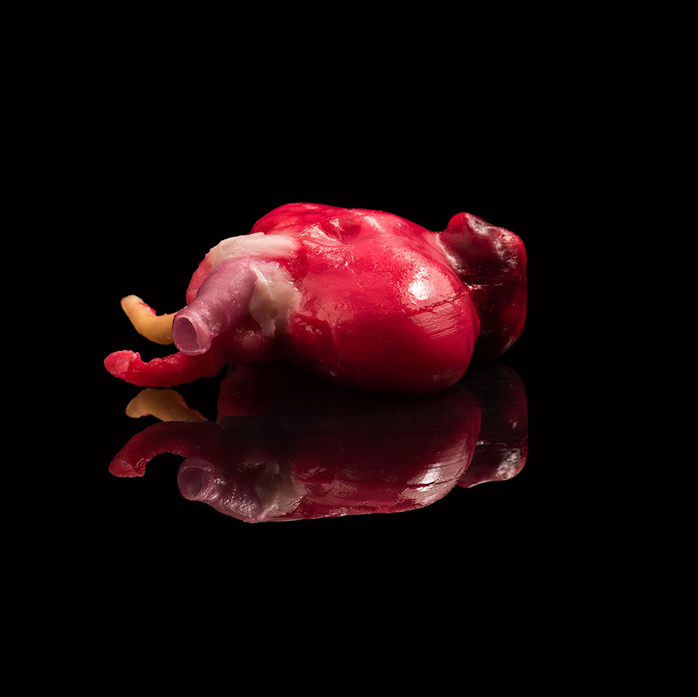
Physicians at the University of Rochester Medical Center have developed a new way to fabricate artificial organs and human anatomy that mimics the real thing, even up to the point of bleeding when cut. These models are able to create highly realistic simulations for training and could soon be widely used to rehearse complex cases prior to surgery.
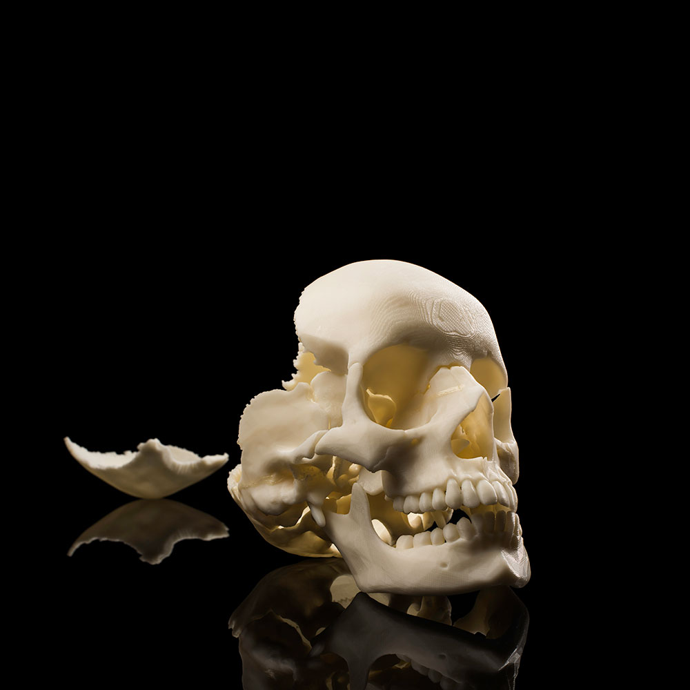
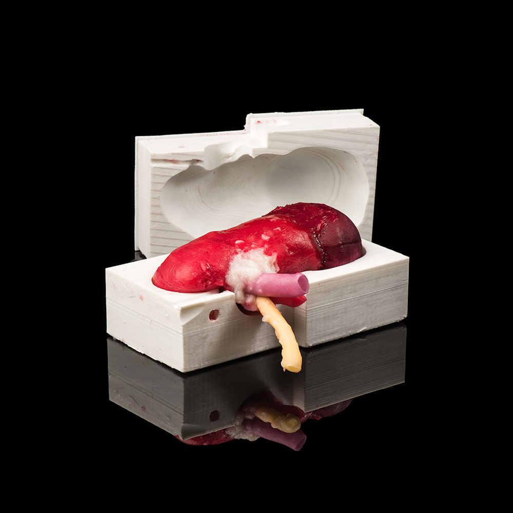
The program, which the creators have dubbed Simulated Inanimate Model for a Physical Learning Experience, or SIMPLE, is the brainchild of Ahmed Ghazi, an assistant professor in the Department of Urology, and Jonathan Stone, a neurosurgery resident who also holds a degree in biomedical engineering. The process entails converting images obtained from medical scans into computer generated designs and, through the assistance of 3D printing, fabricating lifelike organs that can be poked, prodded, and dissected.
“Very few surgical simulations are successful at recreating the live event from the beginning to the end,” said Ghazi. “What we have created is a model that looks, feels, and reacts like a live organ and allows trainees and surgeons to replicate the same experience they would face in the operating room with a real patient.”
Learn more about this 3D printing process >>
University photos / J. Adam Fenster


