New Optica fellow studies bruising associated with abuse, risks of bone fracture, and accumulation of microplastics.
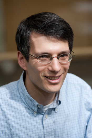
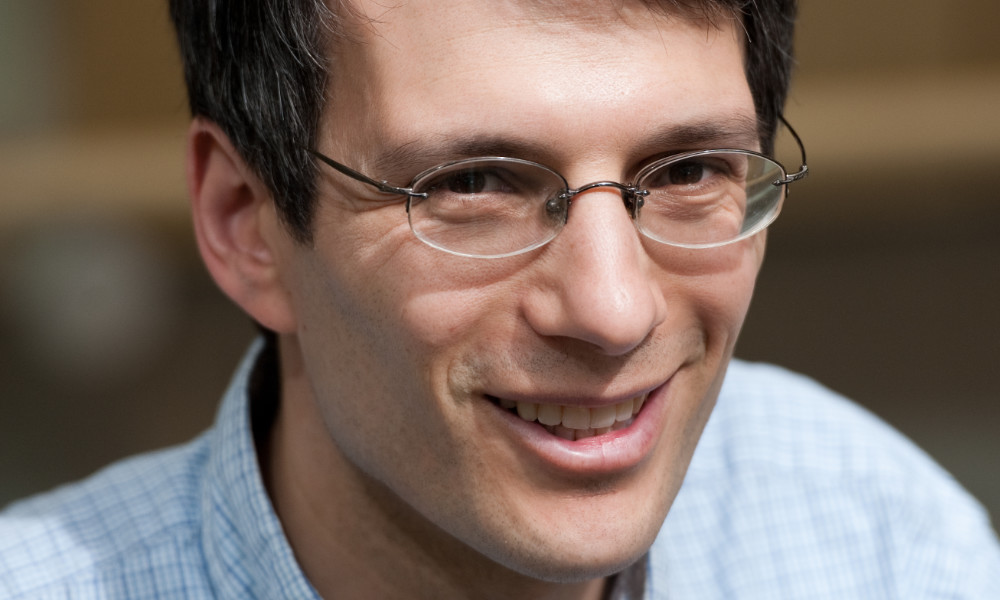
Andrew Berger, a professor of optics at the University of Rochester, scatters light from molecules and cells in novel ways, working with collaborators from many disciplines to detect risk of bone fracture, signs of domestic abuse, and microplastic pollutants in our bodies.
For his achievements, he has been elected a fellow of Optica (formerly OSA), the international society for optics and photonics.
Fellows are Optica members who have served with distinction in the advancement of optics and photonics. No more than 10 percent of the total Optica membership can serve as fellows.
Berger, who joined the University’s Institute of Optics in 2000 and also serves as a professor of biomedical engineering, is being recognized for “significant advances in using intrinsic optical contrast mechanisms to analyze untreated cells and tissues, either in living subjects or in the laboratory.”
Those contrast mechanisms include Raman spectroscopy, in which narrowband lasers scatter light from molecules, revealing detailed information about chemical concentrations. Berger also uses angular scattering of light from single cells to detect changes or differences in the size of the cells’ organelles.
Rochester alumnus also selected
University of Rochester alumnus Rick Plympton ’87 ’99 (MBA), CEO of Optimax Systems Inc., was also elected an Optical fellow for “innovative and outstanding business leadership and service to the Society.” Plympton, who earned his BS at the Institute of Optics, is a member of the Visiting Committee of the Hajim School of Engineering and Applied Sciences.
His recent research projects have included:
- Assessing the risk of bone fracture due to osteoporosis, with Hani Awad, the Donald and Mary Clark Distinguished Professor in Orthopaedics and a professor of biomedical engineering. Initially funded with a University seed grant, the project has generated more than $2.25 million in funding from the National Institutes of Health.
- Using different types of light sources to detect bruising in dark-skinned individuals in order to improve documentation of domestic abuse. The project is a collaboration with researchers at the University’s Susan B. Anthony Center and colleagues at the Institute of Optics.
- Examining the ability of microplastic pollutants to pass through human tissue barriers and accumulate in organs, in collaboration with five other colleagues from the University of Rochester Medical Center, Department of Biomedical Engineering, and the Institute of Optics.
Berger is also an accomplished teacher. He codirects the University’s nano-, bio- and quantum photonics REU (research experience for undergraduates) program and has been recognized with two of the University’s college-wide teaching awards: the Goergen Award for Distinguished Achievement and Artistry in Undergraduate Teaching (2007) and the Edward Peck Curtis Award for Excellence in Undergraduate Teaching (2016).
“I’m always energized by the thought of how I can reach students,” Berger says. “In optics, the challenge of teaching undergraduates is not to be too abstract—to put your energy not into presenting a seamless train of thought but into chopping it up,” he says. So, he uses peer-led workshops, and in lectures checks in with students frequently, using questions, hypothetical scenarios, and other techniques to engage them.
Last year he participated in a University Symposium Series presentation on how faculty members in STEM fields can use online learning tools.
Read more
 Imaging the secret lives of immune cells in the eye
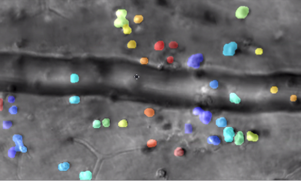
Rochester researchers combine videography and artificial intelligence to track the interactions of microscopic immune cells in a living eye without dyes or damage, a first for imaging science.
 New imaging technology could buy time for pancreatic cancer patients
Tumor shrinkage is one sign of cancer treatment’s efficacy—but Rochester scientists are exploring elasticity and permeability as well.
 Rochester to advance research in biological imaging through new grant
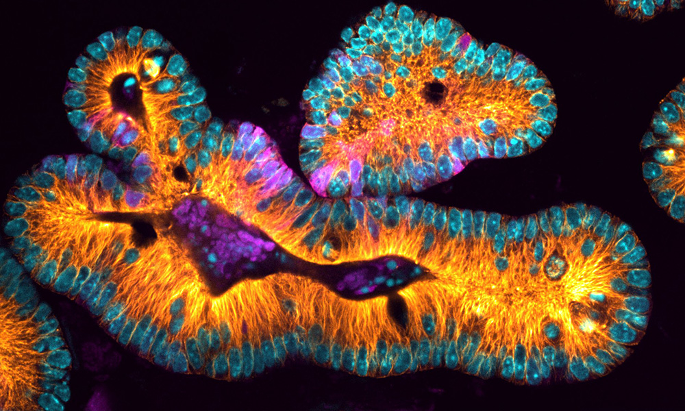
A multidisciplinary collaboration will create a new light-sheet microscope on campus, allowing 3D imaging of complex cellular structures.

