Research
The BSL group has worked on a variety of biomedical topics. Spectroscopy of some type (either wavelength or angular) generally has played a role.
Current front-line projects
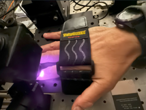
We are working on a light-based method to pre-screen a person for an elevated risk of osteoporosis. By shining 830 nm laser light on a finger as shown below, we are able to obtain Raman (inelastic scattering) spectra from 860-1000 nm. Various peaks in the spectra correspond to minerals (phosphate and carbonate), protein (Type 1 collagen), and lipids. The heights and shapes of these peaks allow us to build machine-learning models that predict bone properties that are typically obtained via X-ray and biomechanical means.
In separate studies, we obtain Raman spectra in vivo on the fingers of human volunteers, in situ on cadaver fingers, and ev vivo on bones extracted from cadavers to compare with the in situ measurement.
Accurately estimating the in vivo spectrum of a finger bone in the presence of overlying soft tissue is a difficult challenge. In our approach, we take measurements at different source-detector separations to obtain spectra with different relative concentrations of bone and soft tissue. We have successfully used libraries of mouse tissues (both tibial bone and various soft tissues) to make accurate estimates. Our cadaver study is in the process of replicating that work with human finger specimens.
In addition to benchtop studies, we are building a cart-based system. By being portable, Raman measurements can be performed at facilities where patients undergo an X-ray technique that is the official standard for osteoporosis diagnosis.
For more information, see the curated list of Raman/bone papers on the Publications page, and check out this poster we made for a 2024 biomedical optics conference.
Under traditional imaging conditions, if a person has darker skin color, a bruise will be harder to detect. All else the same, this leads to inequity in people’s ability to document bodily damage via traditional white-light imaging. This has implications for domestic violence survivors’ ability to trust their lived experiences, as well as to seek help and obtain protection within the legal system.
As part of a multidisciplinary team, we are working to create images of bruises that reduce (ideally eliminate) the effect of baseline skin color. We have developed an imaging system based upon low-cost Raspberry Pi microcontrollers and cameras. By sequentially pulsing millisecond sources at ultraviolet, visible, and near-infrared wavelengths, we can currently obtain a set of images in 1-2 seconds. By summing the images with different weight factors, we hope to produce composite images that reduce the effect of skin color (melanin pigmentation) while preserving the effect of bruising (extra levels of blood or breakdown products).
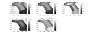
On a technical level, we calibrate to get objective maps of reflectance at each (x,y) pixel. The picture below shows five different grayscale reflectance maps of a volunteer’s arm under different source lighting. The top row shows the red, green, and blue channels of broadband white-LED lighting. The bottom two images are from narrowband LEDs, one blue and the other in the infrared at 950 nm.
For more information, check out this poster from 2024.
Other BSL projects, past and present
[page under development]
[[page under development]]