Researchers have found biometallic interaction on the surface of every daguerreotype they’ve examined with a scanning electron microscope. How and why is this happening? How pervasive and diverse is this condition? What can prevent this from happening?
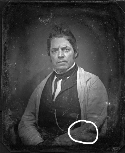
Biometallic interaction on this daguerreotype of an unknown sitter.
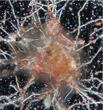
Daguerreotypes exhibit quantum optical characteristics: color in darkfield light microscopy is due to localized plasmon resonance of gold and silver nanoparticles.
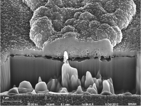
Large image particle in a dark region of the daguerreotype: not the extended diminution of silver at the plating interface below the particle; top surface is gold rich.
A remarkable discovery: the daguerreotype surface engages readily with life forms at the cellular level. It hosts and engages with living organisms and the chemistry of life: Fungi, Bacteria, Plants, Enzymes, Proteins, Nucleic acids, Cytoplasmic fluids.

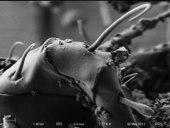
This fade-in is the same particle viewed by SEM at 8K magnification; note the scale bar is 1 micron. This incredible structure is well imaged by SEM because of the nano-metallic infusion into the extra and intra cellular biology, and note the emergence of distinct crystalline structures that have been built and deposited on the exterior. We have no full explanation, nor perhaps does any other branch of science, for all the complex processes that have been at work over the last 160 years in this astounding assembly–which probably still is, or would be progressing, if left undisturbed in its historic petri dish environment.