In Review
 BETTER BONDS: Children improved across a range of developmental measures when their mothers—like Laianna Baker (above)—participated in a Mt. Hope Family Center therapy program, a new study shows. (Photo: Adam Fenster)
Medical Center researchers have discovered that a protein implicated in human longevity may also play a role in restoring hearing after noise exposure. The findings, published in the journal Scientific Reports, could one day provide researchers with new tools to prevent hearing loss.
The study indicates that a gene called Forkhead Box O3 (Foxo3) appears to play a role in protecting outer hair cells in the inner ear from damage. The cells act as a biological sound amplifier and are critical to hearing. But exposure to loud noises stresses them. In some people, the cells are able to recover, but in others the outer hair cells die, permanently impairing hearing. And while hearing aids and other treatments can help recover some range of hearing, there’s currently no biological cure for hearing loss.
“While more than 100 genes have been identified as being involved in childhood hearing loss, little is known about the genes that regulate hearing recovery after noise exposure,” says Patricia White, a research associate professor in the Department of Neuroscience and the lead author of the study. “Our study shows that Foxo3 could play an important role in determining which individuals might be more susceptible to noise-induced hearing loss.”
Foxo3 is known to play an important role in how cells respond to stress. In the cardiovascular system, Foxo3 helps heart cells stay healthy by clearing away debris when the cells are damaged. And people with a genetic mutation that confers higher levels of Foxo3 protein have been shown to live longer.
—Mark Michaud
Treated Mothers Pass Along Benefits of Therapy
Mothers who receive interpersonal psychotherapy after showing signs of major depression fare significantly better than those who receive referrals to other services.
Not only do the moms become better at parenting, according to a new study by researchers at the University’s Mt. Hope Family Center and the University of Minnesota’s Institute of Child Development, but their children also show improvement across a host of important developmental measures. The study was published in the journal Development and Psychopathology.
Children of depressed mothers are at greater risk for a variety of developmental problems, and establishing a secure attachment relationship with a parent is a critical developmental milestone.
Researchers found that after treatment the mothers became better at reading and understanding their toddlers’ temperaments, while the toddlers became less fussy and angry, making them easier to parent.
“It’s a cascading effect for the family,” says lead researcher Elizabeth Handley, a research associate and an assistant professor at the Mt. Hope Family Center.
—Sandra Knispel
‘Hawk Traits’ Foster Kids’ Problem-Solving Skills
How well do standardized cognitive assessments capture children’s cognitive abilities?
Maybe not so well. A new study from the Mt. Hope Family Center suggests that such tests—which don’t consider children’s motivation or environment—may not capture “the specialized repertoire of cognitive skills children in stressful environments have developed as a survival mechanism,” says lead author Jennifer Suor, a doctoral candidate in clinical psychology.
Children growing up in poverty with unengaged caregivers are more likely to do poorly on standardized assessments.
But researchers found that children who at age two showed higher levels of “hawk traits”—heightened levels of aggression, boldness, and dominant behavior in toddlerhood—became better at using problem-solving skills to obtain a blocked reward.
The study, which looked at mothers and their children at ages two and four, was published in the Journal of Child Psychology and Psychiatry.
While on average the kids performed similarly on problem-solving tasks, the children who’d experienced greater caregiving adversity and had more hawk traits were more likely to do better on the problem-solving tasks that involved a reward.
“They were more persistent, tried more solutions, were more engaged,” says study coauthor Melissa Sturge-Apple, an associate professor of psychology and dean of graduate studies in Arts, Sciences & Engineering.
“When kids are faced with poverty and unengaged caregivers, they devote more energy to solving the problem that’s more meaningful to them than one that isn’t,” Sturge-Apple says.
—Sandra Knispel
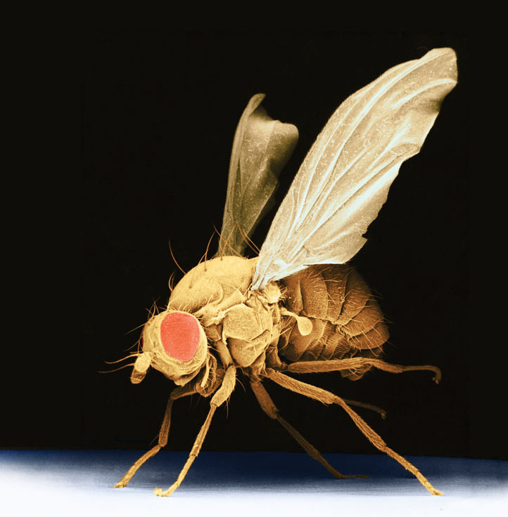
 FOLLOWING ITS GUT: A Rochester study of the intestinal microbiota of fruit flies may have broad implications, given the widespread
use of Drosophila in biologic and genomic studies. (Photo: Science Source)
FOLLOWING ITS GUT: A Rochester study of the intestinal microbiota of fruit flies may have broad implications, given the widespread
use of Drosophila in biologic and genomic studies. (Photo: Science Source)Fruit Flies Offer Gut Check on Bacteria
A Rochester study suggests that researchers may want to rethink common assumptions about laboratory fruit flies—the species of Drosophila melanogaster that’s been a mainstay of biologic and genomic studies.
In the first research to analyze the microbe population found in wild Drosophila, scientists report that fruit flies in the lab may bear little resemblance to what’s seen in fruit flies in the wild—especially when it comes to the bacteria found in their intestinal tracts.
Vincent Martinson, a postdoctoral research associate, led the study, with John Jaenike, a professor of biology, and a Cornell colleague. The findings, published in Ecology Letters, challenge some widely held assumptions about whether an organism’s diet determines the bacteria likely to be found in its gut. The findings also run counter to a recent hypothesis about how the bacterial population should vary among different species.
—Bob Marcotte
Retraining the Brain to See after a Stroke
A kind of physical therapy for the visual system is returning sight to patients who have gone partially blind after experiencing a stroke.
Visual training designed by Rochester researchers was the subject of a new study, published in the journal Neurology. The research offers the first evidence that rigorous visual training recovers basic vision in some stroke patients.
Damage to the brain’s visual cortex prevents visual information from getting to other brain regions that help make sense of it, causing sight loss for up to a half of a person’s normal field of view. Between a quarter of a million and a half of a million people suffer such vision loss each year.
“We are the only people in the U.S. currently using this type of training to recover vision lost after damage to the primary visual cortex,” says senior study author Krystel Huxlin, the James V. Aquavella Professor of Ophthalmology at the Flaum Eye Institute, where she is director of research.
Visual deficits have long been believed to stabilize six months after a visual cortex stroke, and patients are advised to adapt to their vision loss—a marked contrast from treatment for other kinds of strokes. People with stroke damage in areas of the brain that control movement, for example, begin physical therapy as soon as possible and usually recover significant mobility.
Huxlin, who’s also a professor in the departments of neuroscience, brain and cognitive sciences, and the Center for Visual Science, has developed a way of rerouting visual information around the dead areas of the primary visual cortex.
The study also challenges the conventional wisdom that cortically blind patients’ visual deficits stabilize in six months. In the study, the visual deficits of five cortically blind patients who didn’t do visual training got worse, a finding that the team is now verifying with a larger group.
“It might actually be wrong not to train these patients,” says Huxlin. “Our training may be critical for reversing a gradual, very slow, but persistent loss of vision after stroke.” —Susanne Pallo

