
Equipment
SEM/FIB Microscope
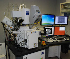
Model:
Zeiss Auriga Scanning Electron Microscope/Focused Ion Beam Tool
Capabilities:
This tool is a high resolution field emission microscope as a well as a lithographic tool using either electrons or Ga+ ions. Sample manipulation is done with a Kleindiek 3-axis probe and Pt deposition is possible using Pt-organic gas injection.
Cost:
Calendar for Reservations:
Specifications:
- Schottky field emission emitter.
- 1 nm resolution in SE mode.
- Four quadrant BSE detector.
- Gas injection system.
- Micromanipulator.
- Up to 30kV Ga+ ion beam.
- Pattern generation systems from both Nabity (NPGS) and Fibics (NPVE).
- EDAX x-ray spectrometer system with mapping capability.
On-line Manual:
SEM systems use charged particles (electrons) to image samples in a vacuum environment. A series of magnetic lenses focuses the electrons to a small point that is scanned, raster fashion. Either reflected (backscattered) electrons or secondary electrons are used to image local variations in samples. X-rays are also generated that are characteristic of the elements present in the sample. Thus, high resolution imaging can be combined with chemical identification. Adding a coincident ion beam column to the system enhances the capabilities to include nano-machining and subsurface analysis.